Introduction to fNIRS
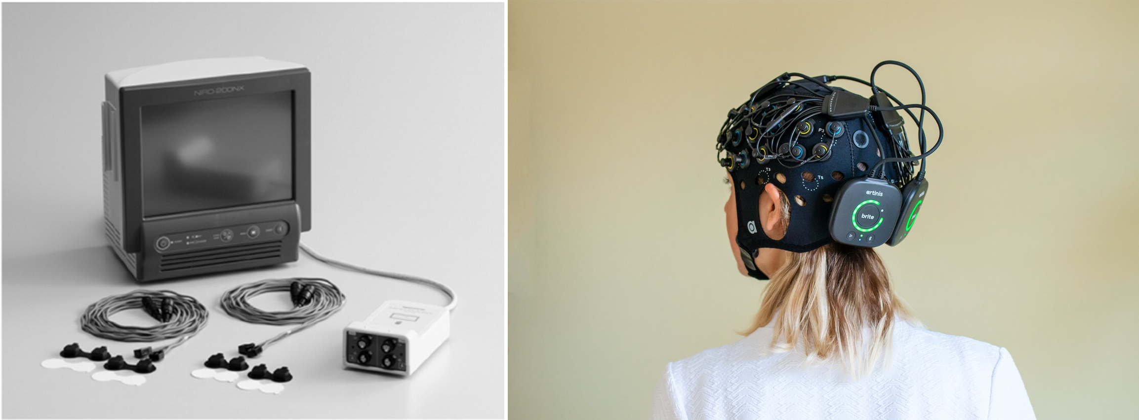
Functional near-infrared spectroscopy (fNIRS), also known as NIRS or optical tomography (OT), is a non-invasive neuroimaging technique increasingly used by the neuroscience community. This method is gaining popularity due to its non-invasive nature, portability, and tolerance for movement during measurements. Compared to other frequently used neuroimaging techniques fNIRS has better spatial resolution than electroencephalography (EEG) and better temporal resolution than functional magnetic resonance imaging (fMRI).
The roots of fNIRS trace back to the 1970s when Frans Jöbsis discovered that biological tissues are relatively transparent to near-infrared light. Building on this intial work, Randall L. Barbour and Ray Aronson introduced the concept of tomographic imaging using near-infrared light in 1988. Barbour and Aronson made fNIRS more accessible and user-friendly for researchers by establishing their company NIRx, allowing it to be integrated into various experiments in neuroscience and other fields.
How does it work?
fNIRS measures changes in brain activity by detecting haemodynamic responses using near-infrared light. Neuronal activation increases local cerebral blood flow (CBF), leading to changes in the concentration of oxygenated haemoglobin (HbO), deoxygenated haemoglobin (HHb), and total haemoglobin (tHb = HbO + HHb). Specifically, when a brain region becomes active, the concentration of HbO increases, while HHb concentration decreases. These changes provide an indirect measure of brain activity, similar to the BOLD (Blood-Oxygen-Level-Dependent) signal used in fMRI.
By utilizing the unique absorption spectra of haemoglobin, which has an absorption window in the near-infrared range (700–1200 nm), fNIRS can detect the relative concentrations of HbO and HHb. Near-infrared light can penetrate several centimeters into biological tissue, allowing the measurement of these haemodynamic changes in the brain’s cortical regions. fNIRS systems consist of light sources and detectors placed on the scalp. The light sources are typically dual-tip LEDs that emit near-infrared light at specific wavelengths, often around 760 nm and 850 nm. These wavelengths are chosen because they are optimal for distinguishing between the absorption spectra of HbO and HHb. The detectors, positioned 3 centimeters from the emitters, measure the amount of light that passes through the brain tissue. The path of the light is not straight but rather follows a “banana-shaped” curve, a phenomenon known as the “photon banana”.
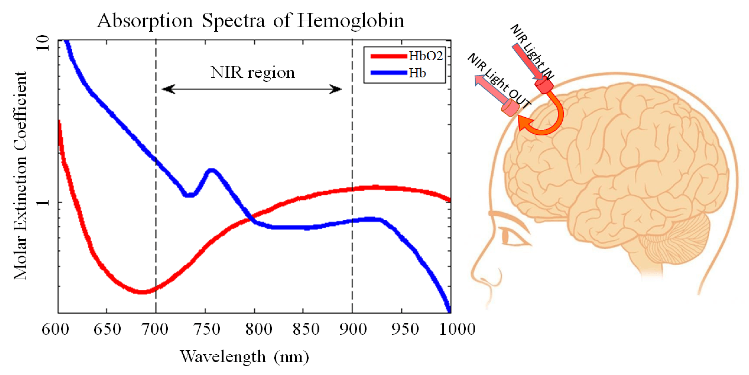
The relationship between light intensity changes and haemoglobin concentrations is described by the Modified Beer-Lambert Law, which accounts for the scattering of photons as they travel through tissue. The modification introduces a differential pathlength factor to adjust for the random travel of photons, providing a more accurate measure of haemoglobin changes.
Learn more about absorbance and transmittance:
NIRS explained: Absorbance & Transmittance by Artinis (2023)
Benefits of fNIRS
- Non-invasive: Unlike some other neuroimaging techniques, fNIRS does not require the insertion of electrodes or exposure to high magnetic fields.
- Affordable and portable: Compared to fMRI, fNIRS systems are more cost-effective and portable, making them accessible for various research settings.
- Experimental flexibility: The portability and ability to use wireless communication with data acquisition systems makes it possible to study participants while they move, opening new possibilities for more naturalistic real-world experiments, increasing the ecological validity of neuroscience research.
- Suitability for specific populations: fNIRS is particularly useful for studying populations such as children, individuals with autism, or other groups that may find traditional neuroimaging methods challenging.
Challenges of fNIRS
- Limited depth penetration: fNIRS is primarily effective for imaging cortical regions, as it can only penetrate about 2–3 centimeters below the scalp.
- Lack of standardization: Being a relatively young field, fNIRS lacks standardized protocols, which can lead to variability in results between studies.
- Challenges with stable contact: Hair can make it difficult to maintain consistent contact between the emitters, detectors, and the scalp, which is affecting data quality.
- Physiological noise and motion artifacts: While fNIRS is more tolerant of movement than some other techniques, it is still vulnerable to noise from physiological sources like heartbeat or breathing, as well as motion artifacts.
Learn more about fNIRS:
fNIRS bootcamp UCLA SCN lab (2019 January)
Introduction to fNIRS webinar by Artinis (2022)
NIRx NIRSport system manual University of Iowa, DEN lab
Functional Near-Infrared Spectroscopy (fNIRS): Principles and Neuroscientific Applications
Interdisciplinary views of fNIRS: Current advancements, equity challenges, and an agenda for future needs of a diverse fNIRS research community
A brief review on the history of human functional near-infrared spectroscopy (fNIRS) development and fields of application
fNIRS course by Prof. Rickson C. Mesquita The present and future use of functional near-infrared spectroscopy (fNIRS) for cognitive neuroscience
Forming research questions
When thinking about possible research questions to be answered with the use of fNIRS first it is important to think about what we can measure with fNIRS. fNIRS measurements indirectly reflect brain activity through the hemodynamic response resolved from detecting changes in the blood oxygenation in cortical areas. With careful considerations and a well-designed study, a wide variety of research questions can be answered, from developmental through more foundational cognitive topics.
Key considerations
Baseline: As fNIRS measures relative changes in haemoglobin concentration, the first important thing to consider is to establish a reliable baseline before presenting stimuli. A good baseline ensures the accuracy of your results and minimizes variability.
Haemodynamic delay: Compared to other methods the haemodynamic response observed with fNIRS is relatively slow, with the change peak happening around 5-10 seconds after stimuli.
Physiological confounds: Physiological factors, such as breathing or heart rate can interfere with fNIRS signals. For instance, Mayer waves (~0.1 Hz) can overlap with brain activity in repeating stimuli which can cause false positive results due to synchronization with the confounds.
Learn more about how to choose a correct baseline:
‘The do’s and don’ts of baselines’ by Artinis
Learn more about how to handle physiological confounds:
‘Best practices for fNIRS publications’ by Yücel et al. (2021)
Confounding effects by SfNIRS
Frequently used experimental designs
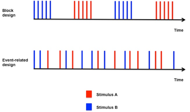
Block design
A block design features alternating blocks of stimuli and rest periods. This approach offers high signal-to-noise ratios, statistical power, and time efficiency. However, careful attention is needed for: - Rest: Allow sufficient time for the response to return to baseline. - Block duration: Avoid overly long blocks to prevent mental fatigue. - Physiological synchronization: Prevent overlap with confounding physiological signals.
Event-related design
In an event-related design, individual stimuli are presented in a randomized or pseudo-random order. This design provides better temporal resolution and lower the anticipatory effect by participants. Important considerations include: - Rest: Keep a minimum of 2 seconds between events. - Analysis: The analysis relies on accurate modelling of overlapping hemodynamic responses, thus the whole analysis model is going to be more complex than with a block design.
General guidelines for experimental design
- Repeat conditions to account for physiological variability.
- Randomize stimuli to reduce anticipatory effects.
- Clearly document the schematic of your experimental design:
- Stimuli, conditions.
- Number and order of blocks or trials.
- Duration of stimuli, rest periods, and intervals.
Learn more about experimental designs and research questions:
‘Which experimental designs to use in fNIRS’ by Artinis
‘fNIRS Experimental Design & Stimulus Presentation’ by Brain Support
UCLA fNIRS Bootcamp Pt. 2 - Study Design Considerations for fNIRS
Ethics: things to consider
Before the research is conducted, the research method has to be submitted to an ethics review board. For example, at Utrecht University the ethics review committee of Utrecht University’s faculty of Social and Behavioural Sciences (FETC) reviews the experimental procedures. Below you will find some things to consider.
Apply with a general research program: One direct and convenient route for ethics approval is to have an fNIRS research program approved. In this research program, the boundary box of the research that will fall under the program is explained. For example, lab-based experiments that investigate individual social cognition or dyadic hyper-scanning. The general procedure is explained as well as the ethical dimension of the research (e.g., risks, max duration, impact on participants).
Provide an fNIRS handout to participants: Within this handout you can explain fNIRS: what is it? How does it work? What are the risks? What will the participant do?
Harm and discomfort to the participant: Because of the non-invasive nature of fNIRS the probability of harm or discomfort is very low. This -as per criteria of the local ethics board- is not “greater than ordinarily encountered in daily life or during the performance of routine physical or psychological examinations or tests”. The duration of the experiment, optode placement, and use of short distance channels, spring holders used, and additional physiological measurement does impact the discomfort. Therefore try to minimise the duration of your experiment as much as possible without compromising the validity of your research.
Exclusion criteria: fNIRS has no exclusion criteria making it extremely suitable to test a diverse population. One important aspect to carefully consider is how hair and skin colour can influence the fNIRS signal and overcome limitations to prevent biases and increase inclusivity of fNIRS research. Accidental findings: Accidental findings are unlikely if not impossible, as fNIRS does not provide structural image of the brain. However, with research progressing this might change.
Anonymity of data: fNIRS data can be stored fully anonymized or pseudo-anonymized.
Optode configuration
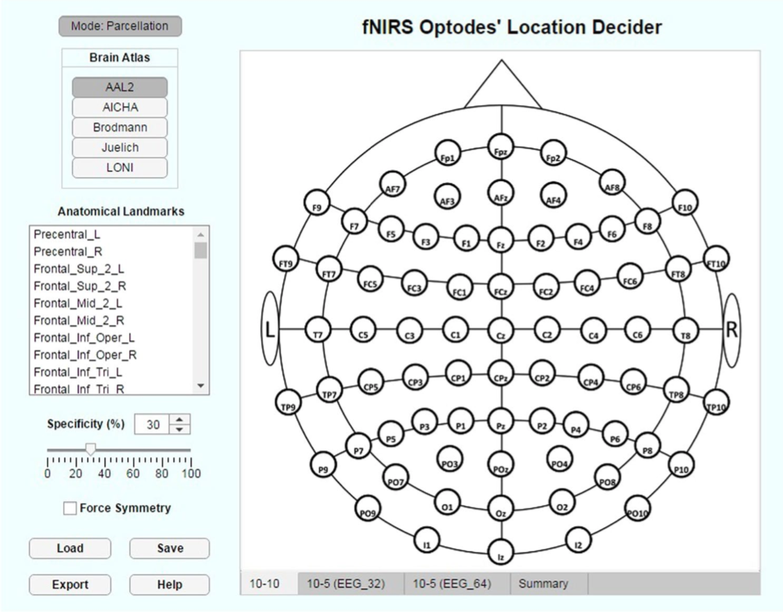
fOLD (fNIRS Optode Location Decider)
According to your brain regions-of-interest, fOLD can automatically suggest an optode configuration.
Reference: Morais, G. A. Z., Balardin, J. B., & Sato, J. R. (2018). fNIRS Optodes’ Location Decider (fOLD): a toolbox for probe arrangement guided by brain regions-of-interest. Scientific Reports, 8(1). https://doi.org/10.1038/s41598-018-21716-z
Installing fOLD: You can download the fOLD toolbox from Github
fOLD demonstration video: https://youtu.be/oA3u8rv5UDQ?si=ZGAVANLH2UGibw1R
Google Slides instruction: https://docs.google.com/presentation/d/1nNQB6xpl4Z1SH9MucbhxNUsTU3D4j-tMUVv-GYJlT08/edit#slide=id.p
Instructions for using fOLD
- Select one of the layouts of which international system you are using for your study (10-10, 10-5 EEG_32 or 10-5 EEG_64).
- Based on the different parcellation atlases, you can choose the anatomical landmarks you are interested in.
- The specificity threshold can be used to define the minimum specificity required for a channel to be included in the optode arrangement based on the selected anatomical landmarks.
- By clicking on the “Summary” tab you can access information about the created optode configuration.
- In case you would like to use a NIfTI file instead of one of the parcellation atlases, you can switch the working mode from “Mode: Parcellation” to “Image Mask”
- Save or export your optode arrangement as ASCII format to import the montage to NIRSite.
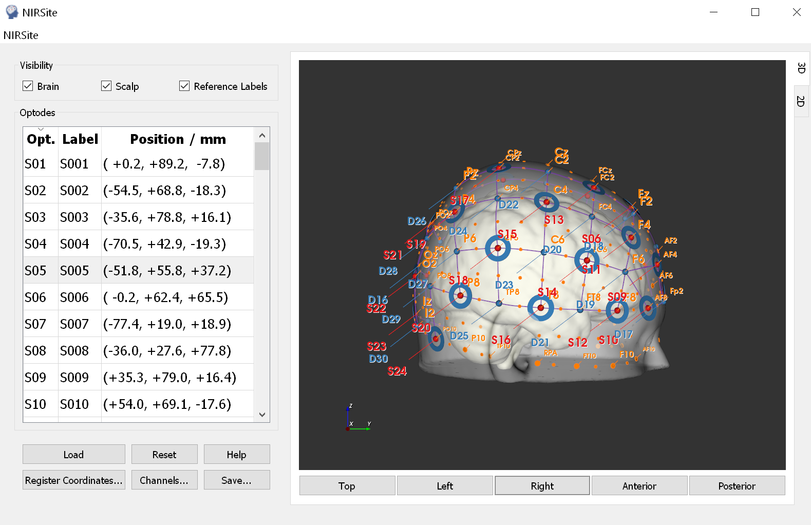
NIRSite
NIRSite allows users to locate optodes onto an MNI space and to input digitized coordinates from other devices into a usable file format for analysis.
Installing NIRSite: You can download the NIRSite software from the NIRX pen drive or from https://support.nirx.de/software. Windows users can download the first available installer (.exe extension). Mac users can download the installer located inside the “Mac” folder (.pkg extension).
NIRSite Getting Started Guide: https://www.nirx.de/downloads/QuickGuides/Updated/Getting%20Started%20-%20NIRSite.pdf
NIRSite User Manual: On the NIRX pen drive or on the NIRX website (https://support.nirx.de/software).
NIRSite Refresher Course video: https://www.youtube.com/watch?v=czk-wZQINNc&t=10s
DEN Lab NIRSite instruction video: https://www.youtube.com/watch?v=DC_H3qLypGM&t=1s
Instructions for using NIRSite
- Select the head model you want to use.
- Creating a new configuration:
- Switch to the 2D view.
- Select which international system you would like to use. The EEG 128 corresponds with the template that comes with the cap.
- Mapping optodes:
- For creating a source: shift + left click (cntrl + left click for Mac), the created optode will appear red.
- For creating a detector: shift + left click (cntrl + left click for Mac) one more time, the created optode will appear blue.
- To create another type of optode: shift + left click (cntrl + left click for Mac) one more time, the created optode will appear green. This ‘other’ optode can be used make it easier to keep track of where other modalities will sit on the head for concurrent recordings, particularly EEG.
- Sources and detectors will automatically form a channel if the channel distance is around 3 cm.
- Keep in mind that sources and detectors will be numbered in the order that they have been created. If you want to renumber the optode you can right click on the optode name in the left panel and then click “Edit”.
- To delete an optode: shift + right click (shift + click on optode, then press delete button and confirm pop up for Mac).
- Once you made your configuration you can switch to 3D view. Use left click + mouse to rotate the 3D template brain.
- Channel distance information can be viewed by clicking on the “Channels” button.
- Mapping short distance channels:
- On the left side panel, click the check box under “Short Ch.” for the sources you want to add a short distance channel to.
- In the drop down under “Bundle” you can select which bundle the short distance channel belongs to.
- Saving montages:
- Name your montage.
- You can upload the configuration directly to Aurora from NIRSite or save it to an external location on your computer first and then upload it. Exporting the file to Aurora will write the montage folder to the default Aurora directory, which is C:~username~(if using OneDrive, it may be written in …~username~…).
- It is recommended to export all the file types.
- Loading a previously created configuration
- Loading an NIRSite file:
- If the previously created file is made for an adult head, select the ICMB-152 head model.
- Click on “load”.
- Select the *_probeInfo.mat file corresponding to your configuration.
- Loading a fOLD created configuration
- If the previously created file is made for an adult head, select the ICMB-152 head model.
- Click on “Import”.
Read more about probe registration:
Probe registration articles by SfNIRS
‘How do you create an fNIRS optodes montage?’ by Cortivision